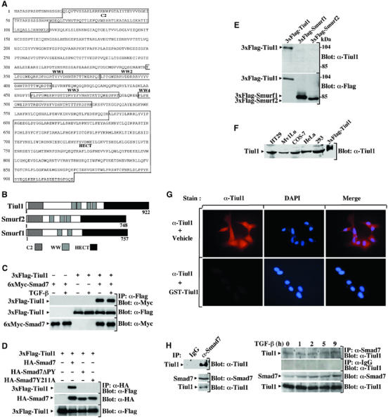
Figure 1.

Smad7 interacts with Tiul1. (A) Amino-acid sequence of the human Tiul1. The conserved domains C2, WW, and HECT of Tiul1 are indicated. (B) Schematic representation of Tiul1, Smurf1, and Smurf2. (C) 293 cells were transfected with 6xMyc-Smad7 in the presence or absence of 3xFlag-Tiul1 and treated with or without TGF-βı (2 ng/ml) for 1 h. Cell lysates were immunoprecipitated (IP) with anti-Flag antibody and immunoblotted (Blot) with anti-Myc antibody. (D) 293 cells were transfected with the indicated combinations of wild-type HA-Smad7, HA-Smad7ΔPY, HA-Smad7Y211A, and 3xFlag-Tiul1. The cell extracts were subjected to anti-HA immunoprecipitation followed by Flag immunoblotting. In each case, the total expression levels of transfected proteins were determined by immunoblotting with the indicated antibodies. (E) Extracts from 293 cells transfected with 3xFlag-Tiul1, 3xFlag-Smurf1, or 3xFlag-Smurf2 were immunoblotted with anti-Tiul1 antibody. The expression of 3xFlag-Tiul1, 3xFlag-Smurf1, and 3xFlag-Smurf2 was monitored by reprobing the same membrane with anti-Flag antibody. (F) Lysates from different cell lines were subjected to immunoblot analysis using anti-Tiul1 antibody. Lysates from 293 cells transfected with 3xFlag-Tiul1 was loaded as a positive control. (G) MDCK cells were stained with anti-Tiul1 antibody and DAPI, and the localization of Tiul1 was visualized under an immunofluorescence microscope. (H) Left panel, COS-7 cell lysates were immunoprecipitated with either normal goat IgG (IP: IgG) or anti-Smad7 antibody (IP: α-Smad7), and immunoblotted with anti-Tiul1 antibody. The total expression of endogenous proteins was monitored by direct immunoblotting. Right panel, Mv1Lu cells were exposed to TGF-βı (2 ng/ml) for the indicated times. In all, 1 mg of total cell lysates was subjected to immunoprecipitation with anti-Smad7 or goat IgG antibodies and immunoblotted with anti-Tiul1 antibody. The total expression of endogenous proteins was monitored by direct immunoblotting.
