
Figure 6.
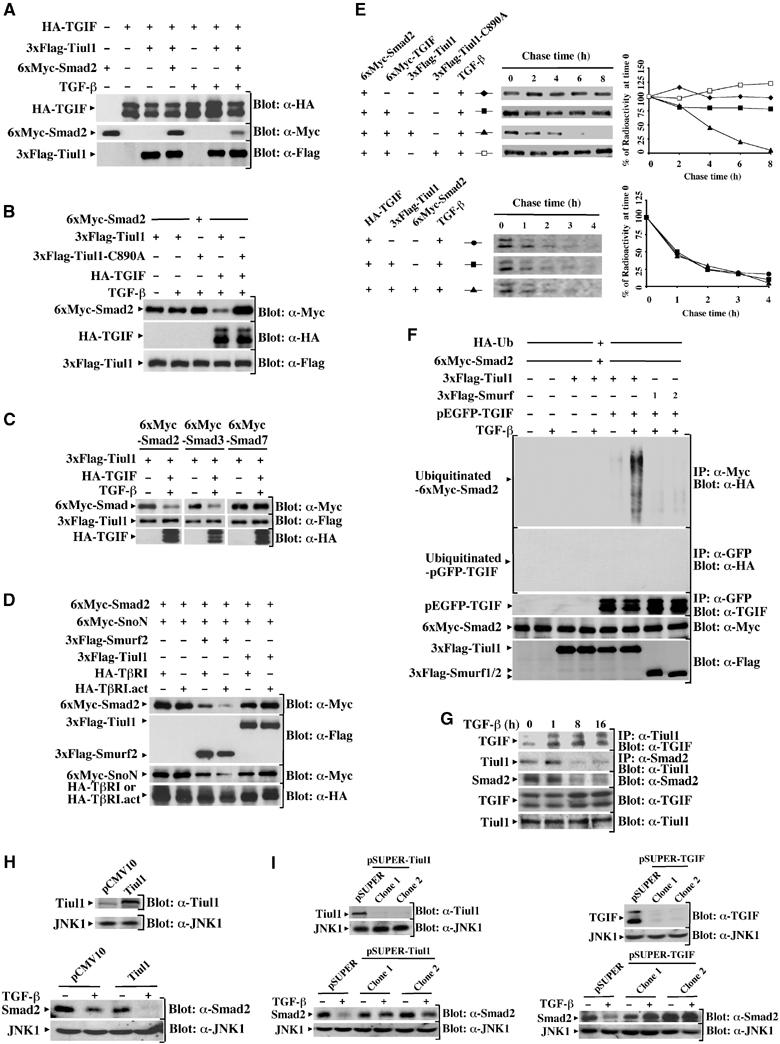
Tiul1 induces ubiquitin-dependent degradation of Smad2 in the presence of TGIF. (A) 293 cells were transfected with the indicated combinations of 3xFlag-Tiul1, HA-TGIF, and 6xMyc-Smad2. Cells were treated with or without TGF-βı for 1 h and cell lysates were subjected to immunoblotting (Blot) with the indicated antibodies. (B) 293 cells were transfected with the indicated combinations of 3xFlag-Tiul1, 3xFlag-Tiul1-C890A, 6xMyc-Smad2, and HA-TGIF. Cells were treated with or without TGF-βı (2 ng/ml) for 1 h and cell lysates were analyzed by immunoblotting with the indicated antibodies. (C) 293 cells were transfected with 3xFlag-Tiul1, HA-TGIF, 6xMyc-Smad2, 6xMyc-Smad3, and 6xMyc-Smad7 as indicated and treated with or without TGF-βı (2 ng/ml) for 1 h. The total expression levels of transfected proteins were determined by immunoblotting with the indicated antibodies. (D) 293 cells were transfected with 3xFlag-Tiul1, 3xFlag-Smurf2, 6xMyc-Smad2, 6xMyc-SnoN, HA-TβRI, and HA-TβRI.act as indicated. The total expression levels of transfected proteins were determined by immunoblotting with the indicated antibodies. (E) 293 cells were transfected with the indicated combinations of 3xFlag-Tiul1, 3xFlag-Tiul1-C890A, 6xMyc-Smad2, 6xMyc-TGIF, and HA-TGIF. Cells were treated with TGF-βı (2 ng/ml) and cell lysates were immunoprecipitated with anti-Myc (Smad2; upper panel) or anti-HA (TGIF; lower panel) antibodies before being visualized and quantified by phosphoimager. Labeled Smad2 (upper panel) or TGIF (lower panel) is plotted at each time point as the percentage of the amount present at time 0. (F) 293 cells were transfected with HA-Ub, together with the indicated combinations of 3xFlag-Tiul1, 3xFlag-Smurf1, 3xFlag-Smurf2, 6xMyc-Smad2, and pEGFP-TGIF. Before lysis, cells were incubated with the proteasome inhibitor MG132 (10 μM) for 6 h. Cells were treated with or without TGF-βı (2 ng/ml) for 1 h and cell lysates were immunoprecipitated with anti-Myc or anti-GFP antibodies before being analyzed by immunoblotting with anti-HA antibody. (G) Mv1Lu cells were treated with TGF-βı (2 ng/ml) for the indicated times and cell extracts were immunoprecipitated with anti-Tiul1 or anti-Smad2 antibodies and immunoblotted with anti-TGIF or anti-Tiul1 antibodies. (H) MDCK cells stably expressing empty vector or 3xFlag-Tiul1 (Blot: α-Tiul1) were treated with or without TGF-βı (2 ng/ml) for 16 h and cell extracts were immunoblotted with anti-Smad2 antibody (Blot: α-Smad2). In all cases, the membranes were reprobed with anti-JNK1 antibody (Blot: α-JNK1) to show that equal amounts of proteins were loaded in the gel. (I) Top, MDCK cells were stably cotransfected with the pEMP4 vector containing a hygromycin resistance gene and empty vector (pSUPER), pSUPER-Tiul1, or pSUPER-TGIF. Clones with reduced expression of Tiul1 or TGIF were identified by immunoblotting total cell lysates with anti-Tiul1 (α-Tiul1; left panel) or anti-TGIF (blot: α-TGIF; right panel) antibodies. Bottom, cells were treated with or without TGF-βı (2 ng/ml) for 16 h and total cell extracts were analyzed by immunoblotting with anti-Smad2 antibody (blot: α-Smad2).