
Figure 7.
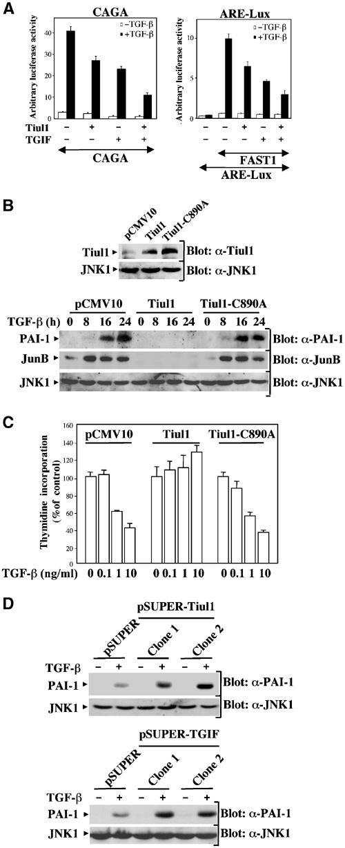
Tiul1 inhibits TGF-β-mediated growth arrest and transcriptional responses. (A) 293 cells were transfected with the reporter constructs (CAGA)9-Lux (left panel) or ARE-Lux, together with FAST1 (right panel) and the indicated combinations of Tiul1 and TGIF. In all cases, cells were treated with (black bars) or without (open bars) TGF-βı (2 ng/ml) for 16 h prior to lysis and then analyzed for luciferase activity. Luciferase activity was normalized to β-galactosidase activity and was expressed as the mean±s.d. of triplicates from a representative experiment performed at least three times. (B) MDCK cells stably expressing the empty vector, 3xFlag-Tiul1, or 3xFlag-Tiul1-C890A (blot: α-Tiul1) were treated with or without TGF-βı (2 ng/ml) for the indicated times and cell extracts were immunoblotted with anti-PAI-1 (blot: α-PAI-1) or anti-JunB (Blot: α-JunB) antibodies. (C) MDCK cells stably expressing empty vector, 3xFlag-Tiul1, or 3xFlag-Tiul1-C890A were treated with the indicated concentrations of TGF-β for 48 h and the rate of cell proliferation was determined by the thymidine incorporation method. Data are expressed as percentages of the relative radioactivity in the absence of TGF-β. (D) Stable MDCK cells with reduced expression of Tiul1 (pSUPER-Tiul1) or TGIF (pSUPER-TGIF) were treated with or without TGF-βı (2 ng/ml) for 16 h and cell extracts were immunoblotted with anti-PAI-1 or anti-JNK1 antibodies.