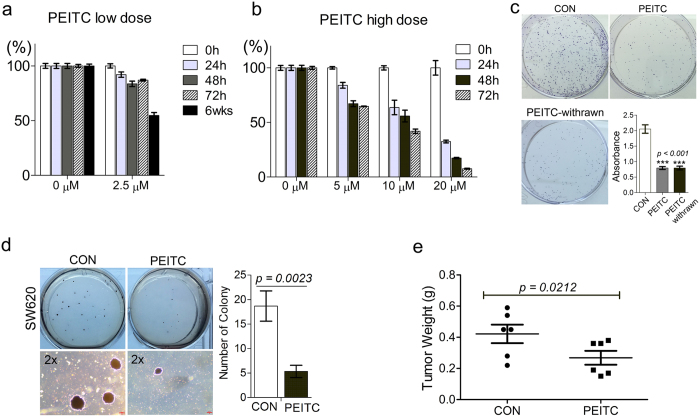
Figure 1. Prolonged exposure to low-dose PEITC reduces growth and tumorigenic potential of colon cancer cells both in vitro and in vivo.
(a,b) MTT assay determination of SW620 cell viability after treatment with low-dose PEITC (a) or high-dose PEITC (b) for the indicated times. Error bars represent mean ± s.d. (c) Colony formation by SW620 cells after 6 weeks treatment with 2.5 μM PEITC (SW620-PEITC) followed by subsequent culture in the presence (PEITC) or absence (PEITC-withrawn) of PEITC treatment for 21 days. SW620 cells exposed to DMSO vehicle only with the corresponding treatment duration served as the control (SW620-CON). Error bars represent mean ± s.d. (d) Anchorage-independent growth of SW620 cells after 6 weeks treatment with 2.5 μM PEITC (PEITC) or DMSO-only vehicle control (CON). The number of colonies formed in soft agar was quantified after 21 days incubation. Error bars represent mean ± s.d. (e) Delayed growth of SW620-PEITC-derived tumors in a mouse xenograft model. SW620 cells were treated with either DMSO only (CON) or 2.5 μM PEITC (PEITC) for 6 weeks prior to s.c. injection into the left or right flank of recipient nude mice. Tumor burden was measured by assessment of gross weight in independent duplicate experiments. Statistical differences between groups were determined using Student’s t-test (mean ± s.d., n = 12, *p = 0.0212).

