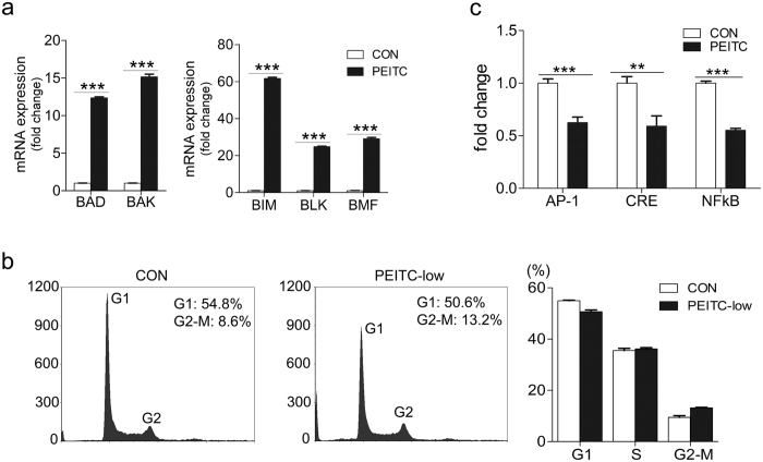
Figure 4. PEITC treatment is associated with tumor upregulation of pro-apoptotic genes.
(a) Up-regulation of pro-apoptotic gene expression in SW620-PEITC cells compared with SW620-CON cells as determined by qRT-PCR. Error bars represent the mean ± s.d., n = 3. (b) Flow-cytometry analysis of propidium iodide-stained SW620 cells treated with 2.5 μM PEITC for 6 weeks (or DMSO-only vehicle control). Shown are representative data from one of three independent experiments conducted. (c) SEAP reporter assay quantification of AP-1, CRE, and NFkB signaling pathway activity in SW620 cells treated with 2.5 μM PEITC or DMSO only (CON) for 6 weeks duration. Error bars represent the mean ± s.d., n = 3. Statistical differences between groups were determined using Student’s t‐test (**p < 0.01; ***p < 0.001).

