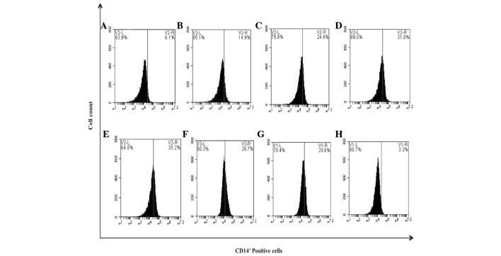
Figure 8.
bDLE induces monocytic differentiation in K562 cells assessed by the expression of the CD14+ surface marker. (A) Untreated cells, (B) 0.07 U/ml bDLE, (C) 0.14 U/ml bDLE, (D) 0.21 U/ml bDLE, (E) 0.28 U/ml bDLE, (F) 0.35 U/ml bDLE, (G) 10 ng/ml phorbol myristate acetate or (H) dimethyl sulfoxide (1.5%, v:v) were incubated for 96 h. Cells were harvested and incubated with anti-CD14 phycoerythrin Texas red in PBS with 1% fetal bovine serum and 0.1% sodium azide for 30 min at 4°C. Samples were washed and resuspended in PBS, and 10,000 events were analyzed by flow cytometry. Flow cytometry data shows representative results from one of three independent experiments. CD, cluster of differentiation; bDLE, bovine dialyzable leukocyte extract.

