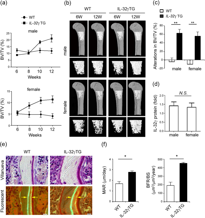
Figure 1. Overexpression of IL-32γ enhances bone volume and bone formation.
(a–c) The femurs from WT and IL-32γ TG male and female mice were isolated at different ages (6, 8, 10, and 12 weeks) and fixed in 4% paraformaldehyde (PFA). The samples were examined by micro-CT imaging. The differences in bone phenotypes between WT and IL-32γ TG mice were analyzed. Bone volume per tissue volume (BV/TV, %) (a), micro-CT images of trabecular bone of femurs from WT and IL-32γ TG mice (b), and alterations (c) were calculated from femur sections using the micro-CT analysis program. (d) Serum IL-32γ levels in female and male TG mice were measured by enzyme-linked immunosorbent assay (ELISA). The results shown are the means ± standard deviation (SD) of 10 mice/group. NS, not significant; **p < 0.01, *p < 0.05. (e,f) Calcein double labeling showing bone formation in 11-week-old WT and IL-32γ TG mice. The mice were injected with calcein twice with an interval of 4 days and sacrificed 2 days after the second injection. The femur bones were embedded, sectioned, and evaluated by Villanueva staining (e). The images were taken using a light microscope and fluorescent light microscope. Scale bar, 10 μm. Histological quantification of mineral apposition rate (MAR, μm/day) and bone formation rate (BFR/BS, μm3/μm2/year) in WT and IL-32γ TG mice (f). The results shown are the means ± SD of 10 mice/group. *p < 0.05.

