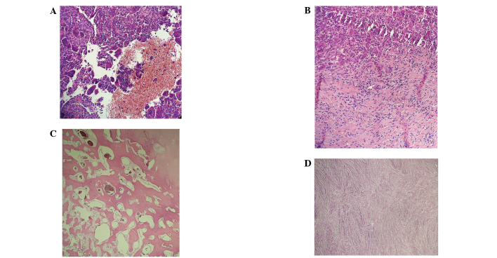
Figure 2.
Biopsy specimens obtained from a 39-year old male with giant cell tumor of bone located in distal meta- and epiphysis of the right femur. (A and B) Prior to treatment abundant hemorrhagic areas, with suspected secondary aneurysmal bone cysts, and foci of fibrosis were evident. Diagnosis was subsequently confirmed by H3F3A mutation testing. (C and D) After 12 months of denosumab therapy, fibrosis and prominent, peripheral ossification of the tumor was identified, indicating a good response to denosumab treatment.

