Figure 1.
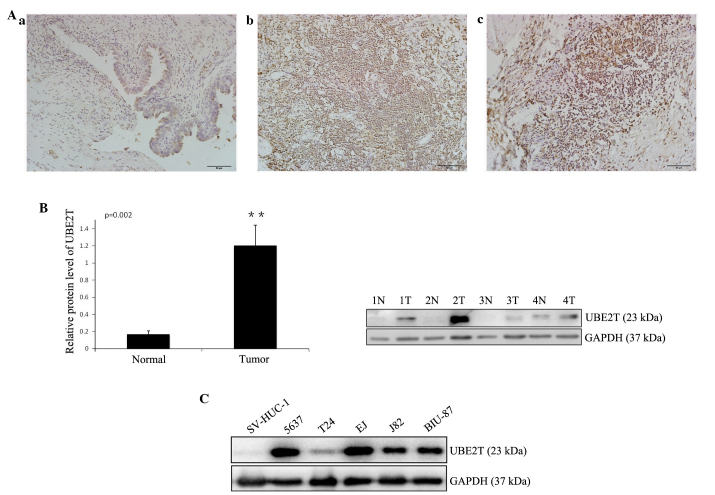
Increased expression of UBE2T in human bladder cancer tissues and cell lines. (A) Representative immunohistochemical staining of UBE2T in normal control tissues and bladder cancer tissues (magnification, ×200; scale bar, 50 µm): (a) Negative or weak UBE2T expression in normal bladder tissues; (b) strong expression of UBE2T in transitional cell carcinoma of the bladder; (c) strong expression of UBE2T in squamous cell carcinoma of the bladder. (B) Representative western blots showing the expression of UBE2T in bladder cancer tissues (tumor/T) and tumor-adjacent bladder tissues (normal/N). The relative UBE2T protein expression is expressed as the mean ± standard deviation from 12 cases, with GAPDH serving as a loading control. **P<0.01. (C) Western blots showing the expression of UBE2T in human bladder cancer cell lines. The normal human urinary tract epithelial cell line SV-HUC-1 was used as negative control. GAPDH served as the loading control. The results represent at least three separate experiments. UBE2T, ubiquitin-conjugating enzyme E2T.

