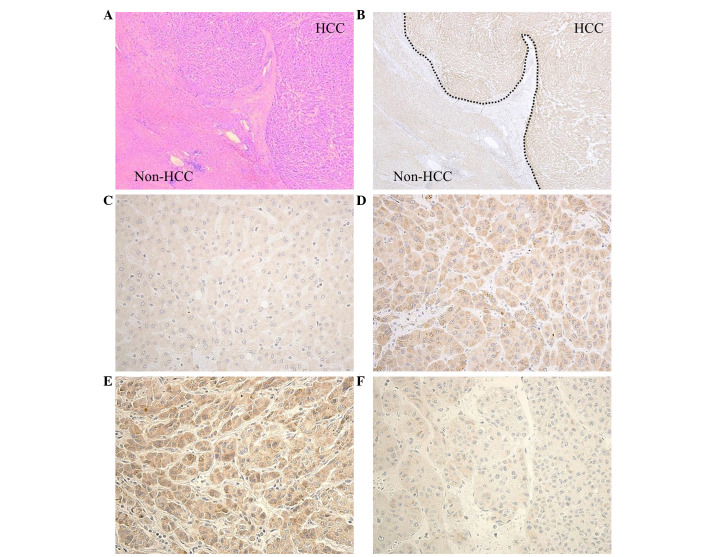
Figure 2.
Expression of PDIA3 in HCC tissue. (A) The representative histology of HCC with trabecular growth pattern and non-HCC tissue. Hematoxylin & eosin staining (magnification, ×40). (B) Intense expression of PDIA3 was observed in the HCC tissue compared with the non-HCC tissue (magnification, ×40). (C) Healthy hepatocytes exhibited only weak PDIA3 staining (magnification, ×400). (D) Clear cytoplasmic staining occurred in HCC cells (magnification, ×400). Representative immunostaining of HCC tissue with (E) high expression and (F) low expression of PDIA3 (magnification, ×400). PDIA3, protein disulfide-isomerase A3; HCC, hepatocellular carcinoma.

