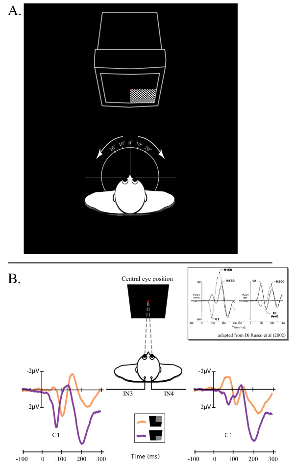
Figure 1.

A. Experimental design. See methods for details. B. Polarity inversion of the C1 component observed on the grand averaged VEP over the 20 subjects and in response to stimuli in the upper (in orange) and lower (in purple) quadrants of the right visual fields. The present polarity inversion was observed on both IN3 and IN4 intermediate occipital sites. The box adapted from Di Russo et al (2002) represents the polarity inversion of the C1 components on the grand average VEPs in response to upper (solid line) and lower (dashed line) hemifield stimuli. In this study, waveforms were collapsed across VEPs to left and right hemifield stimuli and were plotted separately for scalp sites contralateral (left) and ipsilateral (right) to the side of the stimulation. The polarity inversion was observed prominently on occipito-parietal sites PO3/4 using a 10–10-system montage.
