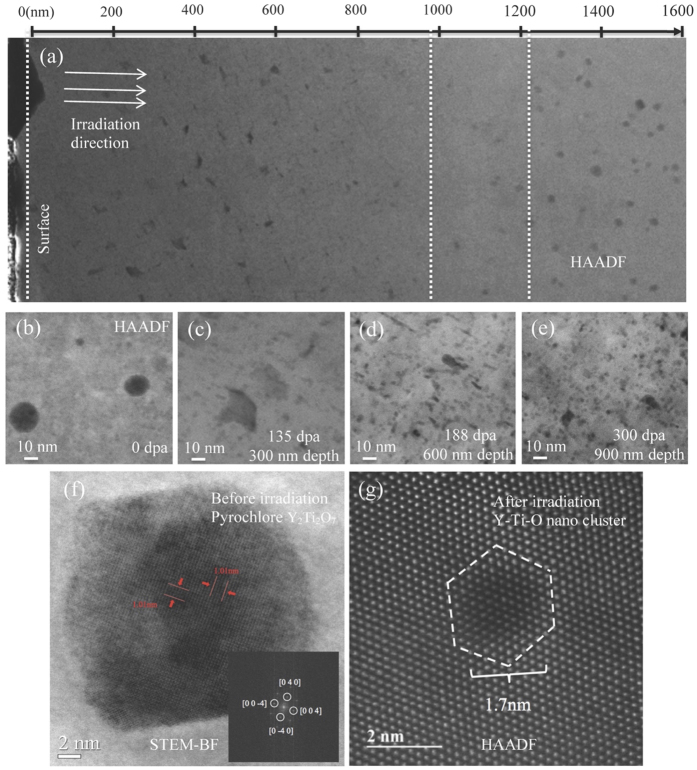
Figure 3. Microstructure changes of COS-2 after heavy ion irradiation.
(a) Cross-sectional HAADF STEM image of a coarse grain in COS-2 after 5 MeV Fe++ irradiation to a fluence of 4.6 × 10¹⁷ ion/cm2 at 460 °C, with 100 appm helium pre-implanted; (b) HAADF image showing oxide distribution in coarse grain of COS-2 before irradiation; (c)~(e) HAADF images showing oxide distributions in various depth with various doses; (f) High resolution STEM-BF image showing the pyrochlore-structured Y2Ti2O7 found in the coarse grains before irradiation; (g) HR-HAADF image showing the newly formed Y-Ti-O nano-cluster in coarse grains after irradiation.

