
Figure 2.
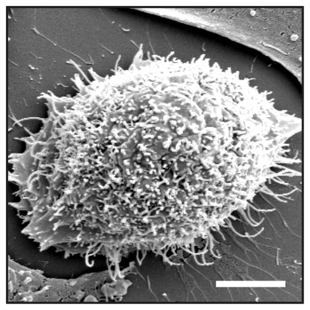
Electron microscopy of HEK 293 Tet-On-FGFRL1ΔC cells. Clone K13ΔC was cultivated in the presence of doxycycline, fixed with glutaraldehyde and prepared for SEM. Inspection by SEM at 10 kV detected numerous filopodia-like spikes with a diameter of ~200 nm. Bar, 5 µm. SEM, scanning electron microscopy.