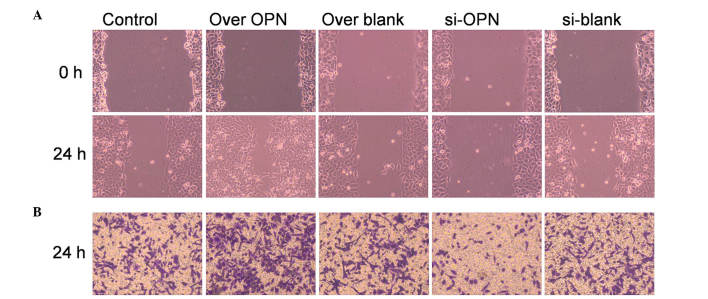
Figure 3.
Effect of OPN on cell migration in the human primary renal cortical endothelial cell line. (A) Wound healing assay was used to examine cell migration. Cells with high OPN expression exhibited stronger ability of healing, since the migrated cell number was significantly higher than that of the control (P=0.0132). OPN-knockdown cells had a lower migrating ability than the control (P=0.0333). (B) Cell invasion was examined by Transwell assay. Cells with high OPN expression displayed stronger invasive ability, since the stained cell number was significantly higher than that of the control (P=0.0223). Cells with low OPN expression (treated by OPN siRNA) displayed a lower invasive ability, and the number of stained cells was significantly lower than that of the control (P=0.0421). Magnification, ×100. OPN, osteopontin; over OPN, over-expression of pcDNA3.1-OPN-green fluorescent protein; over blank, empty carrier over-expression; si-OPN, small interfering RNA interference; si-blank, small interfering RNA.

