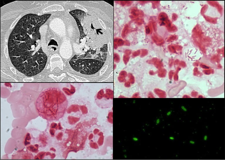
FIG 1.

(Upper left) Section of the chest CT scan, revealing left upper lobe consolidation and cavitation (arrow). (Lower left and upper right) Gram stain of the bronchoalveolar lavage fluid smear. Coloration was performed using a crystal violet-iodine complex and a basic fuchsin counterstain. Magnification, ×1,000. (Lower right) Direct fluorescent antibody stain of the bronchoalveolar lavage fluid using monoclonal antibodies specific for the causative pathogen.
