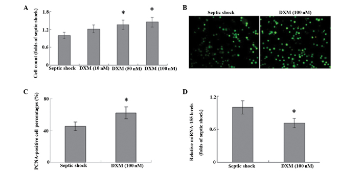
Figure 2.
T cells were isolated from peripheral blood samples and stimulated with 10, 50 or 100 nM DXM for 48 h. Cell proliferation was evaluated using (A) cell count and (B and C) immunofluorescence staining of PCNA (magnification, ×100). (D) miRNA-155 expression levels were detected using reverse transcription-quantitative polymerase chain reaction analysis. *P<0.05 vs. septic shock (control; n=6). DXM dexamethasone; PCNA, proliferating cell nuclear antigen; miRNA, microRNA.

