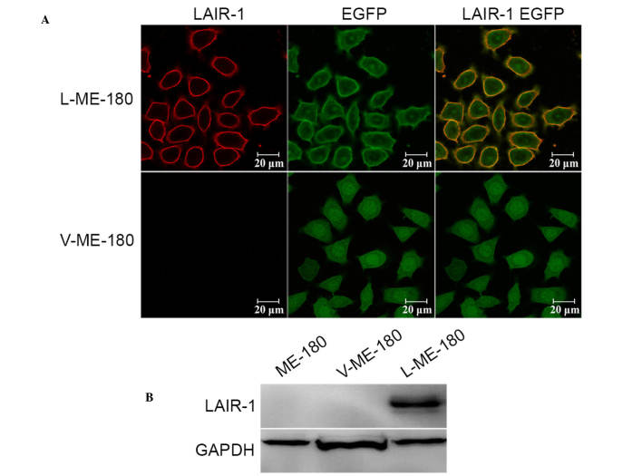
Figure 2.
LAIR-1 expression on ME-180, L-ME-180 and V-ME-180 cells. (A) Laser confocal scanning microscopy was used to analyze LAIR-1 expression. Cells were incubated with Cy3-conjugated anti-LAIR-1 mAb (red). EGFP (green) was expressed in stably transfected cells (L-ME-180) and control cells (V-ME-180). The colocalization of LAIR-1 and EGFP is shown (yellow). (B) Western blotting was used to detect LAIR-1 expression in these cells. GAPDH was used as an internal control. LAIR-1, leukocyte-associated immunoglobulin-like receptor-1; EGFP, enhanced green fluorescent protein.

