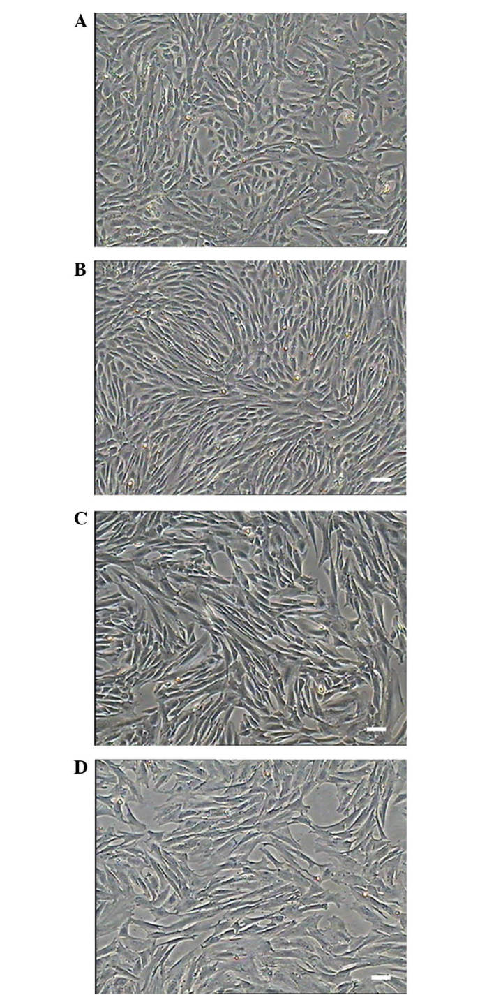
Figure 1.

Morphology of primary cultured and subcultured sheep metanephric mesenchymal stem cells (MMSCs). (A) On day 5 of culture, MMSCs had a distinct ‘shuttle’ shape with clear boundaries and grew slowly. (B) MMSCs at P3. Cells were arranged like fibroblasts and grew in a vortex pattern. (C) MMSCs at P10. (D) MMSCs at P21 appeared senescent (scale bar, 100 µm).
