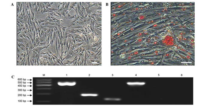
Figure 7.
Adipogenic differentiation of sheep metanephric mesenchymal stem cells (MMSCs). (A) As a negative control, cells cultured in complete medium showed no changes in morphology and were negative for Oil Red O staining. (B) After induction for 12 days, MMSCs became fibroblast-like to oblate and formed numerous intracellular lipid droplets. Lipid droplets were stained with Oil Red O (scale bar, 100 µm). (C) Expression of adipocyte-specific genes LPL and PPARG were detected by reverse transcription-quantitative polymerase chain reaction in the induced group after induction for 12 days. Adipocyte-specific genes were not expressed in the control group. Lane 1: GAPDH served as the internal control in the inducted group; lane 2: PPARG was positive in the inducted group; lane 3: LPL was positive in the inducted group; lane 4: GAPDH served as the internal control in the control group; lane 5: PPARG was negative in the control group; lane 6: LPL was negative in the control group.

