Figure 4.

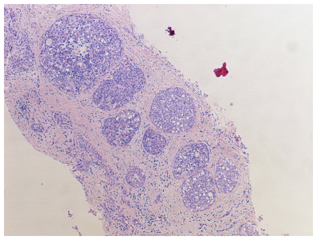
Core biopsy showing expanded acini filled with a pleomorphic proliferation of cells. Haematoxylin and eosin, ×40 magnification.

Core biopsy showing expanded acini filled with a pleomorphic proliferation of cells. Haematoxylin and eosin, ×40 magnification.