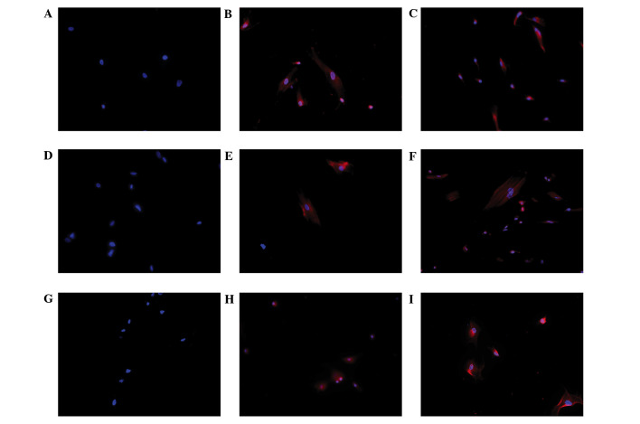
Figure 5.
Immunofluorescence staining of Leydig cell differentiation of human umbilical cord mesenchymal stem cells (HUMSCs) induced in DIM and LC-CM. HUMSCs induced by DIM and LC-CM were positively stained for CYP11A1, CYPY17A1 and 3β-HSD. Immunofluorescence for 3β-HSD was (A) negative in control cells and positive in (B) LC-CM and (C) DIM-induced cells. Blue, nuclei; red, 3β-HSD in cytoplasm. Immunofluorescence for CYP11A1 was (D) negative in control cells and positive in (E) LC-CM and (F) DIM-induced cells: Blue, nuclei; red CYP11A1 in cytoplasm. Immunofluorescence for CYP17A1 was (G) negative in control cells and positive in (H) LC-CM and (I) DIM-induced cells: Blue, nuclei; red, CYP17A1 in cytoplasm. (Scale bar, 100 µm.) CM, conditioned medium; DIM, differentiation-inducing medium; 3β-HSD, 3β-hydroxysteroid dehydrogenase.

