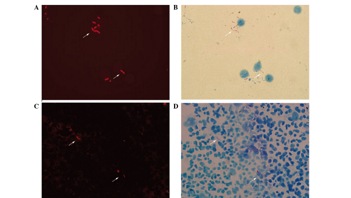
Figure 1.
Micrographs of acid-fast bacilli obtained with fluorescence microscopy and transmitted light microscopy (modified Z-N staining). The fuchsin-stained acid-fast bacilli revealed (A) bright orange-red fluorescing rods (shown by arrows) under fluorescence and (B) red, lightly curved rods (shown by arrows) under transmitted light (magnification, ×1,000). These rods were also visible (shown by arrows) by (C) fluorescence and (D) transmitted light microscopy at a lower magnification (×800).

