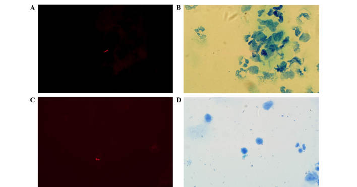
Figure 2.
Comparison of the morphologic changes prior to and following treatment (modified Z-N staining; magnification, ×1,000). The shapes of the acid-fast bacilli were diverse and appeared thin and slightly curved prior to treatment, and are shown (A) under fluorescence and (B) under transmitted light. However, the bacilli became shorter and thicker following treatment, as shown (C) under fluorescence and (D) under transmitted light.

