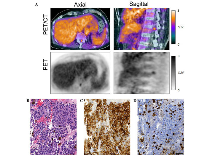
Figure 1.
(A) 18F-fluoro-ethyl-tyrosine PET with and without CT scans showing marked elevation of intramedullary tracer uptake at the 12th thoracic vertebra. (B-D) Histomorphological and immunohistochemical findings. (B) Hematoxylin and eosin staining revealed a tumor with increased cellularity, cytological atypia and cytoplasmic as well as extracellular melanin pigment. The cytomorphological aspect was dominated by epithelioid cells arranged in nests or fascicles. Mitotic figures were observed. (C) Immunohistochemical examinations revealed strong immunoreactivity for HMB45. (D) ~10% of the tumor cell nuclei expressed the proliferation-associated antigen Ki-67 (MIB-1; scale bar, 50 µm). The lesion was classified as melanocytic tumor with increased proliferative activity, compatible with a malignant melanoma. PET, positron emission tomography; CT, computed tomography; SUV, standardized uptake value.

