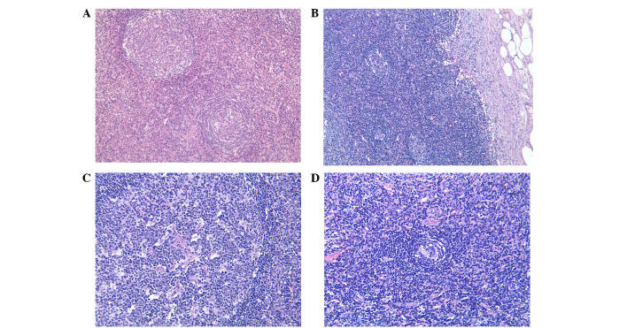
Figure 3.
Histological examination of the left inguinal lymph node biopsy specimen before treatment. (A) Hematoxylin and eosin staining showing a marked diffuse proliferation of large atypical lymphoid cells and lymphoplasmacytic infiltration with irregular fibrosis. (B) Marked thickening of lymph node capsule. (C) Obliterating phlebitis. (D) Reduced area of germinal centers. Magnification, ×100.

