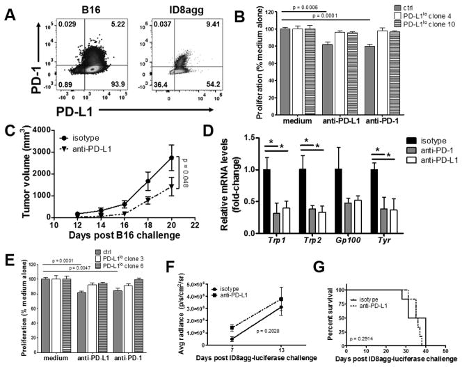
Figure 2. αPD-L1 reduces B16 growth and metastatic spread in NSG mice.
A. PD-1 and PD-L1 expression in B16 melanoma and ID8agg ovarian cells measured by flow cytometry. B. Proliferation in vitro of B16 cells ± αPD-L1 or αPD-1 (50 μg/mL each) determined by MTT versus control (ctrl, set at 100%). p-value, unpaired t test. C. NSG mice challenged with indicated B16 cells and treated with αPD-L1 200 μg every other day starting one day following challenge. p-value, two-way ANOVA. D. qPCR for indicated genes from whole lung lysates from mouse challenged as in C, given αPD-L1 or αPD-1 200 μg every other day starting on day following challenge, day 18. Unpaired t test. *, p < 0.05, **, p < 0.01. E. Proliferation in vitro of ID8agg cells treated as in B. NSG mice challenged with ID8agg-luciferase and treated with αPD-L1 200 μg every other day starting one day following challenge. P values for average luciferase radiance (F) by two-way ANOVA and for survival (G) by log-rank test.

