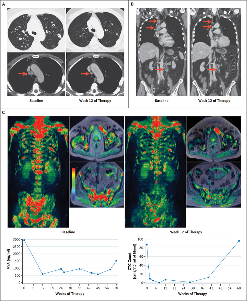
Figure 3. Radiologic Evidence of Tumor Responses to Olaparib at Week 12.
Panel A shows CT scans of the chest, obtained in the lung and soft-tissue window settings, from a 61-year-old man with metastatic, castration-resistant prostate cancer (Patient 39) who had a response to olaparib; there was shrinkage of the lung and nodal (arrows) metastatic deposits after 12 weeks of therapy (right), as compared with baseline (left). Whole-exome sequencing showed a somatic homozygous deletion of BRCA2. Panel B shows CT scans with coronal reconstruction in a 70-year-old man with a somatic BRCA2 frameshift insertion (p.Y2154fs*21) and somatic deletion of the second allele (Patient 20). The scans show the response in the mediastinal and abdominal lymph nodes (arrows). The patient received treatment for a total of 48 weeks. Panel C shows multiparametric whole-body MRI scans, including diffusion-weighted imaging, with coronal three-dimensional reconstruction and selected axial images in a 79-year-old man (Patient 1) who had a response to olaparib, with an 85% reduction in the PSA level. The patient received treatment for a total of 73 weeks. The images show reduction in the water content within the skeletal metastasis, which in conjunction with other findings on imaging would be consistent with tumor regression during therapy (right), as compared with baseline (left). Next-generation sequencing of the baseline bone marrow–biopsy specimen revealed a somatic missense mutation within the ATM phosphoinositide 3-kinase catalytic domain (p.N2875S), with no evidence of genomic loss of the second allele and with maintenance of ATM expression on immunohistochemical assessment.

