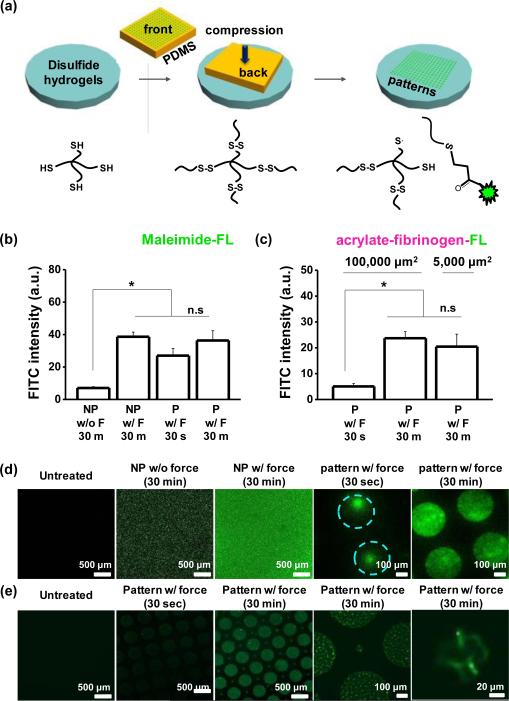
Fig. 3.
(a) Schematic illustrating the process used to pattern different molecules via compression on disulfide hydrogels. Fluorescence intensity for patterned (P) and non-patterned (NP) gels with (b) fluorescein-5-maleimide and (c) acrylate fibrinogen fluorescein with or without force and different holding time. Representative laser scanning confocal microscopen images of disulfide hydrogels when patterned with (d) fluorescein-5-maleimide and (e) acrylate fibrinogen fluorescein. (*P<0.05, t-test)

