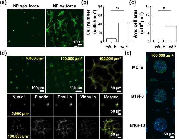
Fig. 4.
(a) Representative images of MSCs on the unpatterned gel-protein substrate with or without compression. Plot of (b) cell number and (c) average cell area differences with or without compression. (d) Representative immunofluorescence images of MSCs captured on protein patterned islands (100,000 or 5,000 μm2) and im-munostained with Paxillin and Vinculin. (e) Immunofluorescence images of different types of patterned cells (MEFs, B16F0, and B16F10) cultured on protein conjugated disulfide hydrogels. (*P<0.05, **P<0.05, t-test)

