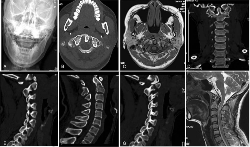
Figure 1.

Open mouth view X-ray showed lateral displacement of C1 lateral mass which indicting C1 unstable fracture; CT showed multiple fragments of the axis (an unstable odontoid fracture type IIA, traumatic spondylolisthesis of C2-C3, and a C1 burst fracture); MRI showed type IIA transverse atlantal ligament rupture of C1 and an unstable C2-C3.
