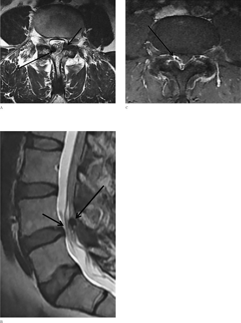
Figure 1.
A) Axial T2-weighted MRI of the lumbosacral (LS) spine at L4-L5 level demonstrates a hypointense lesion in right lateral epidural space (long arrow) causing mass effect on the thecal sac and a smaller cyst (short arrow) in left epidural space. B) Sagittal T2-weighted image of the LS spine demonstrates crowding of the cauda equina spinal nerve roots (short arrow) by the epidural lesion. C) Axial post-contrast fat-saturated T1-weighted MRI showing enhancement of the wall of the right epidural gas cyst and adjacent thickened thecal sac (arrows).

