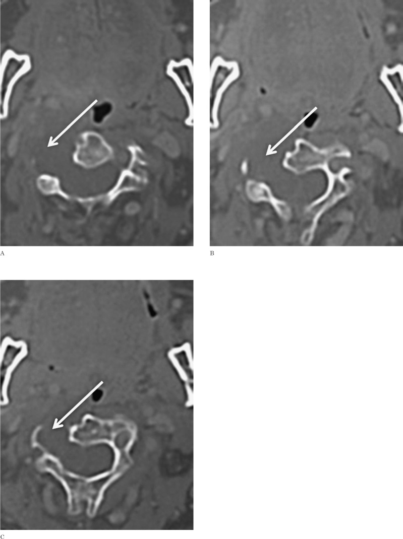
Figure 1.
CT study. Axial CT images after intravenous administration of contrast material show a destructive hypodense lesion, with no apparent enhancement. The lesion causes markedly lytic destruction of the right lateral aspect of the C2 and C3 vertebral bodies and of the right transverse processes, extending into the spinal canal through a very enlarged intervertebral C2-C3 foramen and causing cord compression.

