Figure 2.
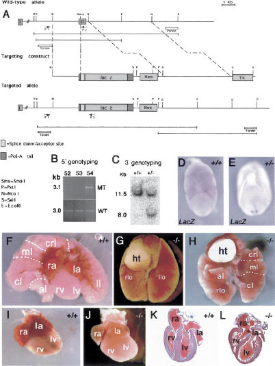
Targeted inactivation of cerl-2 gene. (A) Schematic representation of the wild-type cerl-2 locus, targeting vector, and targeted allele. The positions of primers, restriction enzyme sites, and the probe used for PCR and Southern blot analysis, respectively, are shown. (B) PCR-based 5′-genotyping of wild-type and targeted ES cell DNA. (C) Genomic Southern blot of PstI-digested tail DNA, prepared from newborn offspring of a mating of heterozygous mice. (D,E) LacZ in situ hybridization in wild-type (D) and heterozygous (E) embryos. (F-H) Thoracic organs of newborn wild-type (F) and cerl-2-/- littermates displaying left lung isomerism (G) or inverted situs (H). (I,J) Hearts of newborn wild-type and cerl-2-/- littermates, respectively. (K,L) Frontal sections of the hearts depicted in I and J, respectively, showing the atrial septal defects (asterisk) in cerl-2-/-. (al) Accessory lobe; (cl) caudal lobe; (crl) cranial lobe; (ht) heart; (ml) middle lobe; (la) left atrium; (llo) left lobe; (lv) left ventricle; (ra) right atrium; (rlo) right lobe; (rv) right ventricle.

