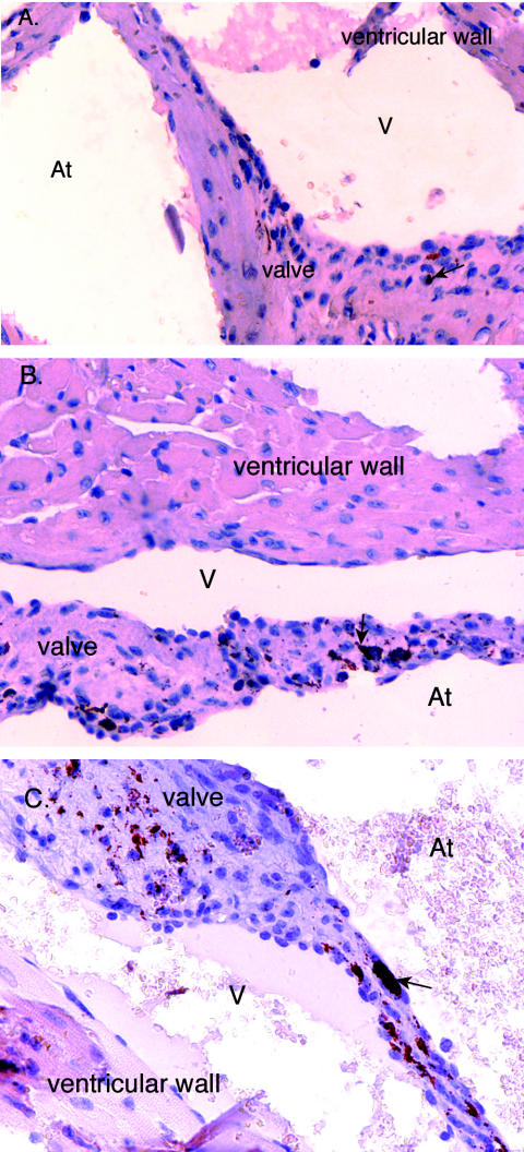
FIG. 6.
Immunolocalization of C. burnetii in cardiac valve sections from mice challenged with 107 C. burnetii organisms at 28 days postinfection. C. burnetii was detected by the ABC method (Vector Laboratories). (A) Valve section from wild-type mouse; (B) valve section from iNOS−/− mouse; (C) valve section from p47phox−/− mouse. The red to rust-colored deposits represent the presence of C. burnetii (arrows denote examples). At, atrial side; V, ventricular side. Magnification, ×40.

