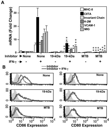
FIG. 3.
M. tuberculosis and 19-kDa lipoprotein inhibit IFN-γ-induced expression of molecules involved in MHC-II Ag processing and presentation. Macrophages were infected with M. tuberculosis (MOI = 30:1) or incubated with 30 nM 19-kDa lipoprotein for 24 h. IFN-γ (2 ng/ml) was added for 24 h. (A) RNA was isolated, and quantitative real-time RT-PCR was used to analyze expression of MHC-II, CIITA, invariant chain, H-2M, VCAM-1, and MIG. The results are expressed as mean ± the standard deviation for triplicate samples. Welch's t test was used to determine the significance of the difference in expression of the indicated mRNA species in the presence of IFN-γ alone versus expression with IFN-γ plus M. tuberculosis or 19-kDa lipoprotein. A single asterisk indicates a P value of <0.05, and two asterisks indicate a P value of <0.01. (B) Macrophages were analyzed for expression of CD86 (left panels) and CD80 (right panels) by flow cytometry. Shaded histograms denote isotype-matched negative control antibody labeling, thin lines indicate expression in the presence of the inhibitor indicated in the panel (none, 19-kDa lipoprotein, or M. tuberculosis), and thick lines indicate expression with both inhibitor and IFN-γ. The percent control expression (expression with IFN-γ alone) was calculated based on specific mean fluorescence values (MFVs), i.e., specific MFV = MFVspecific antibody − MFVisotype-matched control antibody) by using the following equation: (specific MFVInhibitor + IFN-γ/specific MFVIFN-γ) × 100. IFN-γ-induced expression of CD86 was reduced to 50 and 28% of control by M. tuberculosis and 19-kDa lipoprotein, respectively.

