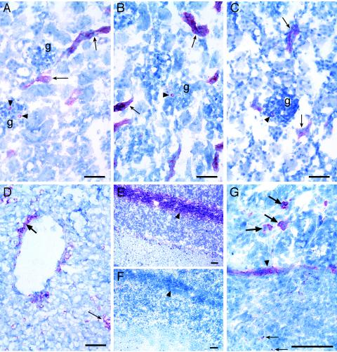
FIG. 1.
Immunohistochemical identification of Stx binding sites and Gb3 in neonatal porcine tissues. Anti-CD77/Gb3 (A), Stx1 (B), and Stx2 (C) binding to tubules (arrows) and glomeruli (arrowheads) in sections of the kidney (g, glomerulus) are shown. (D) Stx2 binding to endothelial cells lining a vessel (thick arrow) and sinusoidal cells within the parenchyma (thin arrow) of the liver. Anti-CD77/Gb3 (E), but not Stx1 (F), binding to nerve fibrils in the white matter (arrowheads) of the cerebellum was observed. (G) Stx2 binding within a Peyer's patch (thin arrow), to smooth muscle (arrowhead), and to cells within the villous lamina propria (thick arrows) in the ileum. Digital images were captured directly with a Digital Spot RT Slider camera (Diagnostic Instruments, Inc., Sterling Heights, Mich.) using MetaVue, version 5.0.7, imaging software (Universal Imaging Corp., Downingtown, Pa.). Bar = 50 μm.

