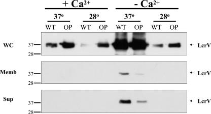
FIG. 1.
The high-temperature and low-calcium dependence of LcrV synthesis is relaxed in Dam-overproducing Y. pseudotuberculosis. Whole-cell (WC), membrane (Memb), and supernatant (Sup) fractions (12) were prepared from wild-type (WT) and Dam-overproducing (OP) Y. pseudotuberculosis grown under the indicated conditions according to methods described previously (12, 44, 45). For each growth condition, total protein extracts corresponding to 2.0 × 10⁶ cells (∼20 μg of protein/well) were subjected to sodium dodecyl sulfate-polyacrylamide gel electrophoresis, transferred to a polyvinylidene difluoride membrane (Pierce), and probed with mouse anti-LcrV monoclonal antibodies (1:4,000 dilution). Peroxidase-conjugated sheep anti-mouse immunoglobulin G (1:40,000 dilution; Amersham Biosciences), was used as the secondary antibody, and hybridization was detected by chemiluminescence using Supersignal West Femto Maximum Sensitivity Substrate (Pierce) followed by a 2-min exposure to film. Inspection of corresponding Coomassie-stained gels showed similar band intensities of nonregulated proteins under all conditions tested (data not shown). Western analysis of lcrV+ and lcrV− strains confirmed that the 37-kDa protein was LcrV (data not shown).

