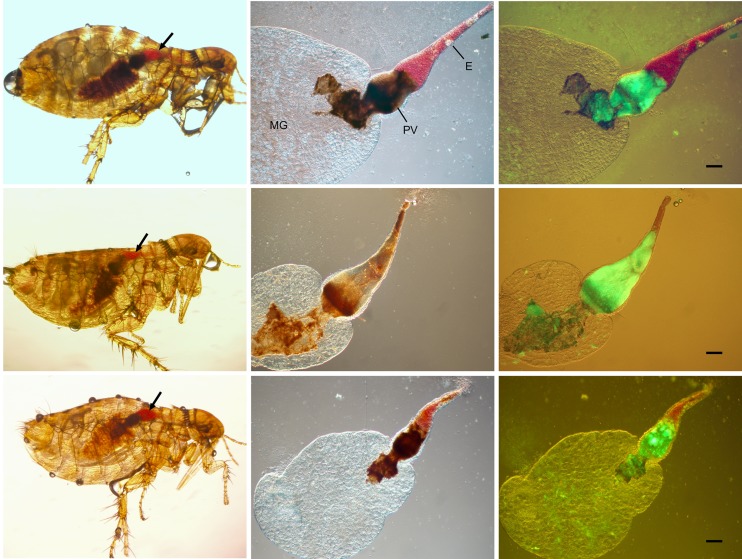
Fig 2. Y. pestis causes complete proventricular blockage in O. montana fleas.
The left panels show the typical blockage phenotype in O. montana fleas immediately after a feeding attempt. Fresh red blood (arrows) that was unable to enter the midgut (MG) is seen in the esophagus (E). The fleas were infected with GFP-expressing Y. pestis. The middle and right panels are images of the dissected digestive tracts from the same fleas visualized by DIC and a combination of DIC + fluorescence microscopy, respectively, and confirm the presence of a dense Y. pestis biofilm that completely fills and blocks the proventriculus (PV). Scale bars = 100 μm.

