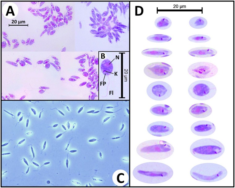
Fig 3. Morphology of trypanosomatid cells in axenic cultures.
(A) Photomicrographs of Leishman stained Zelonia australiensis promastigotes cultured in M3, viewed under oil emersion microscopy (1000X magnification). (B) Photomicrograph of a round promastigote with gross morphological characteristics indicated including the nulcleus (N), kinetoplast (K), flagellar pocket (FP), and flagellum (Fl). (C) Wet mount photomicrograph of live axenically cultured Zelonia australiensis promastigotes viewed under phase contrast microscopy (400X magnification) showing several forms. (D) Photomicrographs of the various Z. australiensis forms as seen in Leishman stained slides, prepared from axenically cultured parasites. The parasite shows a high degree of pleomorphism in culture. This has been reported for other trypanosomatids, and limits the use of morphology for classification of these organisms [16, 101].

