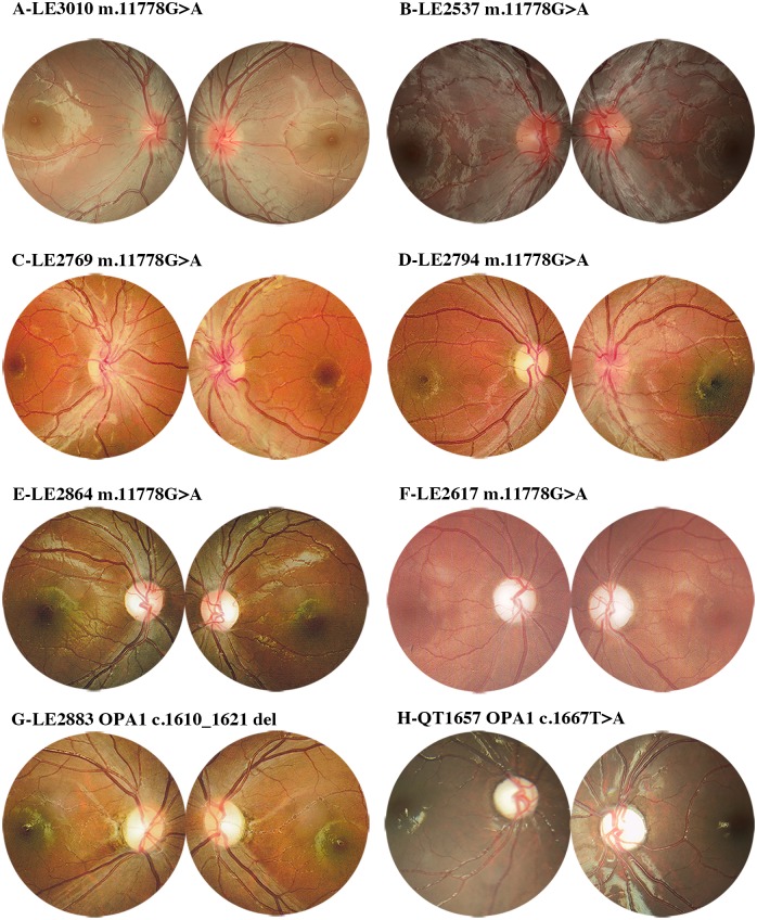
Fig 2. Fundus manifestations of patients with DOA and LHON.
(A-F) LHON-A (edema of optic nerve) patients manifested as: (A) bilateral optic nerve edema without telangiectatic vessels; (B) bilateral optic nerve hyperaemia with telangiectatic vessels and microvascular tortuosity (C) temporal optic pallor with vascular tortuosity in both eyes; (D) temporal optic pallor with microvascular tortuosity in the right eye and optic nerve hyperaemia with telangiectatic vessels in left eye. LHON-SP manifested as: (E) temporal optic atrophy without changes in the vessels in both eyes; (F) diffused atrophy of the optic disk in both eyes. (G-H) The manifestations of DOA (G-H) were similar to LHON-SP with temporal optic atrophy (E, G) or diffused optic pallor (F, H). The patients IDs and mutations are located above the fundus images.

