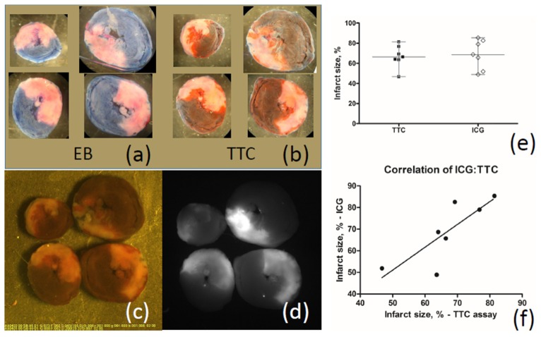
Fig. 4.
Comparison of images of infarction areas obtained by the new method of visualization using ICG and using the traditional method of staining with Evans blue and TTC. There was no difference in infarct size determined by two methods of staining. (a), (b) - Transverse heart slices stained with Evans blue and TTC after 120 min of reperfusion; (c), (d) – The images of the same slices obtained using ICG-scope television camera: (c) - in the visible light, (d) - in the NIR light; (e) – Comparing infarct size evaluated by ICG fluorescence and TTC staining. Medians are not different; (f) – Correlation of infarct sizes obtained by ICG fluorescence and TTC staining is significant.

