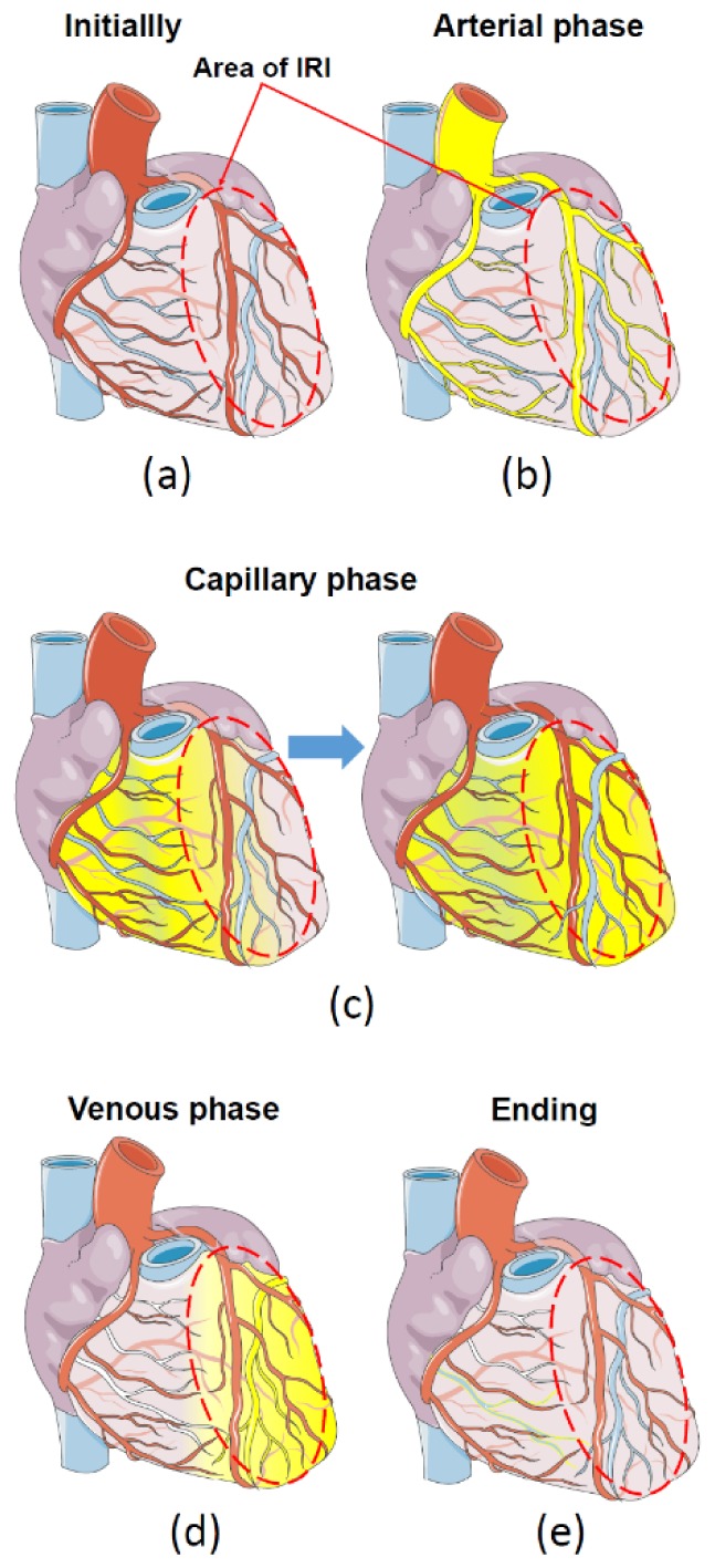
Fig. 5.

Phases of distribution of fluorescent dye (ICG) in the heart with regional IRI. (a) - initially, before administration of ICG, red oval denotes the area of IRI. (b) - the first, arterial phase - distribution of ICG on large coronary vessels. (c) - Capillary phase - distribution of ICG on small vessels. (d) – the venous phase of ICG with delay of ICG in the area of IRI and complete clearance from undamaged myocardium, (e) - the final washout of ICG.
