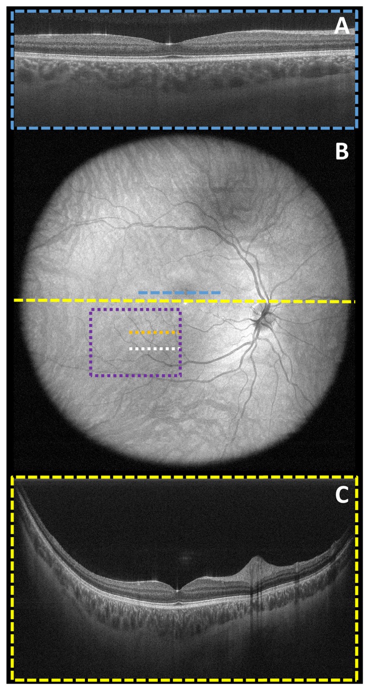
Fig. 5.

Demonstration of WF-OCT system’s large (70° or ~22 mm x 22 mm) FOV and uniform intensity distribution in a healthy volunteer. No WSAO correction was applied in these images. A 5x averaged foveal B-scan (A) with the deformable mirror in the flat position (AO off) was shown (blue dashed line) in context of the un-averaged wide field-of-view SVP (B), which depicted the imaging range of the wide-field sample arm. A 5x averaged wide-field B-scan (C) taken across the horizontal meridian (yellow dashed line) demonstrated good choroidal penetration even in the periphery of the retina. The orange and white dotted lines correspond to the locations of the independently acquired B-scan images shown in Figs. 6 and 8, while the purple dotted box corresponds to the independently acquired volume image of Fig. 7.
