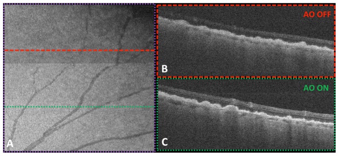
Fig. 11.
Maximum intensity projection (A) of peripheral (27°) vasculature when the deformable mirror was switched between the unoptimized (top) and optimized (bottom) mirror shapes. WSAO increased the brightness of the en face volume projection (~4.4 mm x 4.4 mm) by 26.3%. Corresponding B-scans for regions without (B) and with (C) wavefront correction were shown on the right. The volume image was acquired in the location indicated by the purple dotted box in Fig. 10.

