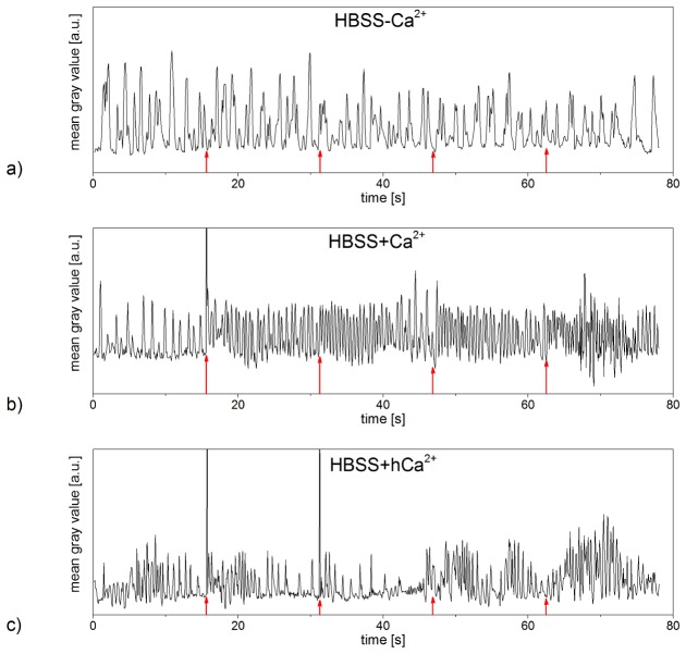
Fig. 4.
Contractions of neonatal rat cardiomyocytes pre and post laser stimulation with a radiant exposure of 8 mJ/cm2 and an irradiation time of 10 ms. While the laser stimulation did not affect the contractile behavior of the examined cardiomyocyte in HBSS-Ca2+ (a), it induced significant increases of the contraction frequency in cardiomyocytes in HBSS + Ca2+ (b) (referring to Visualization 2 (13.6MB, MP4) ). Cardiomyocytes in HBSS + hCa2+ exhibited higher contraction frequencies after laser treatment as well (c) (referring to Visualization 3 (13.6MB, MP4) ). Laser irradiation occurred every 15.6 s (red arrows), every peak represents one contraction of the examined cardiomyocyte. Data shown represents the exemplary contractile behavior of one single out of at least n = 7 examined cells or set of cells in HBSS-Ca2+, HBSS + Ca2+ and HBSS + hCa2+, respectively. Narrow peaks are laser light detected by the camera if it was coincident with the frame acquisition.

