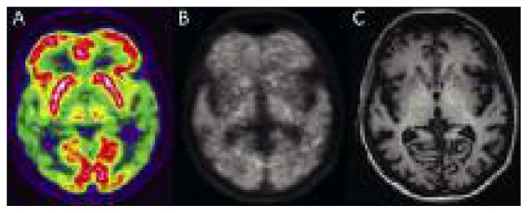
Figure 7.
Images of a patient with mild dementia.
(A) [18F]FDG, (B) [18F]florbetaben, and (C) T1-weighted MPRAGE MRI images show severe bilateral reduction of glucose metabolism in both temporoparietal regions, associated with diffuse deposition of amyloid-seeking tracer in the gray matter. MRI shows mild enlargement of cortical sulci. (Image courtesy of Prof. Diego Cecchin, Department of Medicine (DIMED), Nuclear Medicine, University of Padua, Italy).

