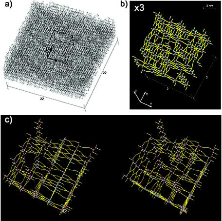
FIG. 4.
Simulated tertiary structure of the staphylococcal murein. The chain length distribution corresponds to the high-performance liquid chromatography data observed for S. aureus (5). The normalized cross-linking degree is 83%. All glycan strands are oriented perpendicularly to the plasma membrane, with GlcpNAc termini pointing upward. (a) Panorama of a simulated murein matrix occupying 22 by 22 positions for the strands and 45 layers thick. Dashed lines outline a cube corresponding to a smaller murein fragment that is selected for enlargement in panel b. Boldfaced arrows correspond to three coordinate axes. (b) Top view of the threefold-enlarged fragment of the murein, composed of 5 by 5 glycan strand positions and 20 levels thick, extracted from the matrix as outlined by dashed lines in panel a. The z axis is perpendicular to the plasma membrane, which is represented by a black background. The residues of GlcpNAc are presented as red dots, while those of MurpNAc are presented as cyan blue dots. Cross-linked oligopeptide chains are clearly visible as yellow zigzags. (c) Stereo view of the simulated murein matrix shown in panel b.

