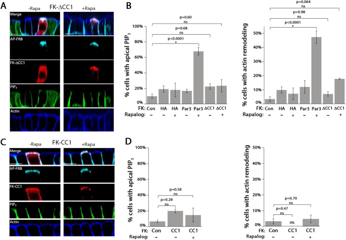
FIGURE 3:
The CC1 region of Par3 is necessary but not sufficient to drive apical membrane remodeling. (A) Confocal XZ-slices of MDCK cells expressing PH-Akt-GFP (PIP3; green) transfected with AP-FRB and FK-∆CC1 (red; HA) and treated with vehicle (–Rapa) or 200 nM Rapalog (+Rapa) for 60 min. Actin was visualized with phalloidin staining (blue). (B) Quantitation of the appearance of apical PIP3 and actin rearrangement in cells transfected with the indicated constructs in the presence or absence of Rapalog. (C) Confocal XZ-slices of cells expressing FK-CC1. (D) Quantitation of the appearance of apical PIP3 and actin rearrangement in cells transfected with the indicated constructs in the presence or absence of Rapalog. Error bars, SEM. The p value was determined by one-way ANOVA followed by post hoc Tukey’s test. n = 3. Scale bar, 10 µm.

