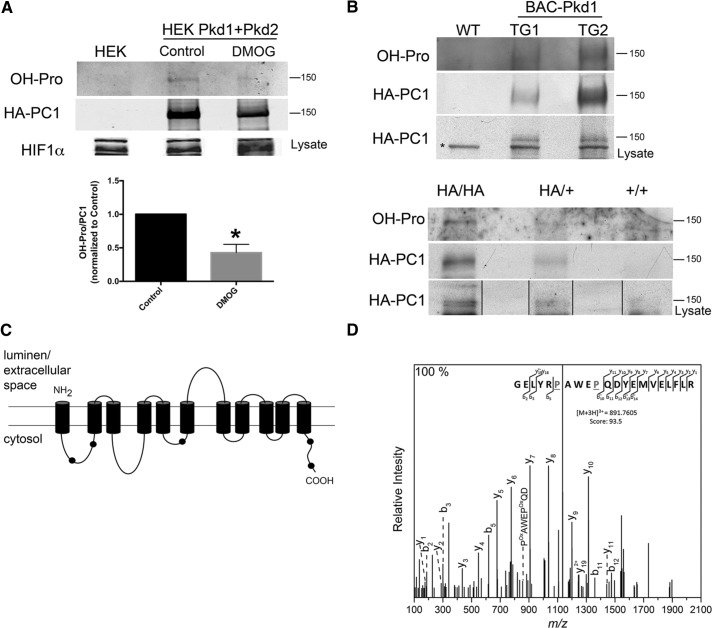
FIGURE 2:
PC1 proline hydroxylation. (A) Western blot analysis to detect the presence of OH-Pro on PC1 that was immunoprecipitated from HEK293 cells that overexpress PC1 and PC2 and were untreated (Control) or treated with 1 mM DMOG for 48 h; the graph shows the quantification of three independent experiments; *p < 0.05. (B) Western blot analysis of OH-Pro on PC1 that was immunoprecipitated from kidneys of mice expressing 3xHA-tagged Pkd1 (top; kidneys from two different transgenic animals, TG1 and TG2, and one wild-type mouse [WT]). The asterisk indicates a nonspecific band recognized by the anti-HA antibody in mouse tissue. Western blot analysis of OH-Pro on PC1 immunoprecipitated from kidneys of heterozygous (HA/+) and homozygous (HA/HA) HA-tagged PC1 knock-in mice; material from a wild type mouse is also shown (bottom). (C) Schematic representation of the PC1 CTF, showing the localization of the peptides containing OH-Pro that were identified by MS (black dots). The position and sequence of the peptides corresponding to each black dot are presented in Supplementary Table S1. (D) Tandem MS of the peptide GELYRPAWEPQDYEMVELFLR from PC1 confirming the presence of two OH-Pro residues (P). Characteristic b and y ions and internal ions were annotated for clarity.

