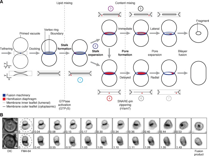
FIGURE 1:
Fragment formation during vacuolar lysosome fusion. (A) Working model describing how intralumenal membrane fragments form during homotypic vacuolar lysosome membrane fusion. Numbers indicate reaction intermediates that were visualized using TEM and are shown in Figure 5D. (B) Live yeast cells stained with FM4-64 to label vacuole membranes were imaged using HILO microscopy. Examples of vacuole fusion events are shown as a series of inverted micrographic images acquired over time (minutes). Dashed lines outline each cell observed by differential interference contrast. Scale bars, 1 µm.

